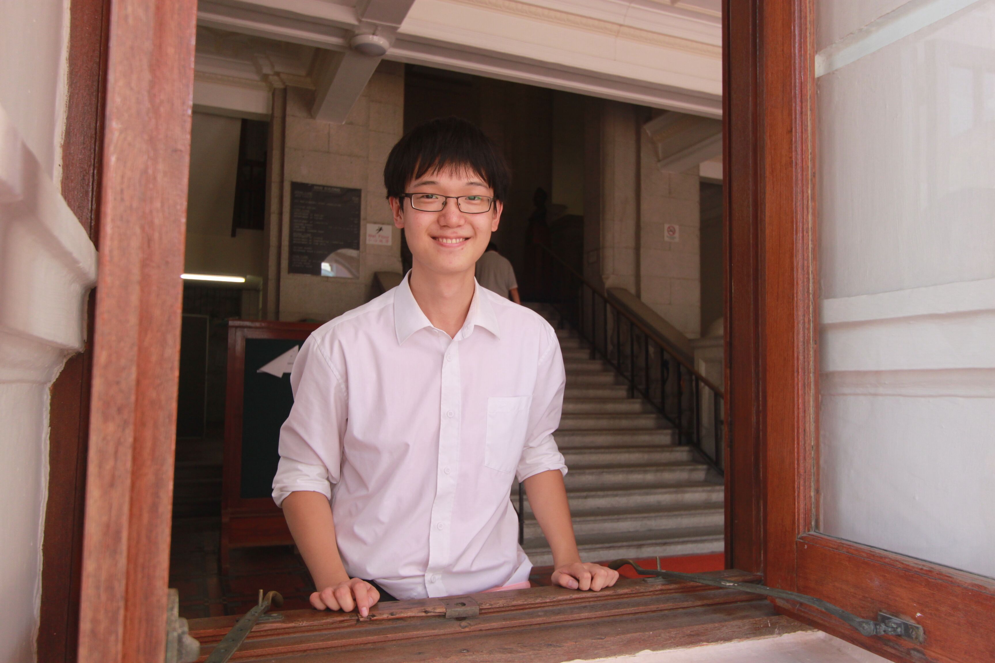
It all started with two suitcases.
When he first came to the Cambridge Stem Cell Institute in 2013, Dr. Bon-Kyoung Koo left behind all the samples at his previous lab in Utrecht, Netherlands. As one of the youngest principal investigators in this world-renowned Institute, he brought with him no more than what a college freshman would bring to her orientation day.
But he had something very special with him. He was one of the first scientists to be involved in the development of a new technique that grows mouse intestinal tissues in a petri dish, and he brought the protocol to Cambridge, England. “It is a very efficient and economical model system to study genetics,” said Dr. Koo, one word at a time, as if he wanted to make sure I understood.
“…a simple but powerful tool to extract stem cells and their progeny from mouse small intestines and retain their physiology.”
Biologists rely on specialized tools to carry out experiments and test their hypotheses. Tissue culture, for example, allows biologists to study the behavior of mammalian cells in the laboratory. However, traditional cell culture protocols are often insufficient to faithfully recapitulate delicate biological structures outside of animal bodies. The intestinal organoid culture technique pioneered by Dr. Koo and his colleagues comes to the conventional methods’ rescue. This new technique is not just a variation of the established protocols that can make cells live longer in the dish; it is a simple but powerful tool to extract stem cells and their progeny from mouse small intestines and retain their physiology for further study of their functions.
Basically, all you need to do is to harvest small intestines from genetically modified mice with genes that you’re interested in, gently scrape the intestine with a small slide commonly used in the lab for microscopes, cut it with scissors to small pieces of a few millimeters thickness to expose the inner wall of the intestine, and wash the intestine pieces with common buffer solutions by vigorously shaking the tube. Cells from the remaining fragment, once transferred to the dish with the right medium and antibiotics, can reform their characteristic finger-like protrusions (called “villus”) and depressions (called “crypt”) structure and grow for as long as six months. In other words, in just about 3 days, you can mass produce these small intestine-like (thus the name “organoid”) structures that exhibit the same functionalities as if they were in living mice.
Scientists have long been aware of the presence of villus and crypt structures on the inner surface of mammalian intestines. We know that these structures can increase the surface area of the intestine, thus promoting the absorption of nutrients into the body. But what drives those intestinal cells to form such fine structures as the villi and crypts, and why do some of these villi end up developing into tumors? With the highest self-renewal rate among all mammalian tissues, intestinal cells have a lifetime of about only 5 days and can become cancerous relatively easily, which is why colorectal cancer is one of the most common types of cancer in the entire Western world, affecting more than 130,000 Americans each year, according to the National Cancer Institute.
Dr. Koo is addressing this puzzle with the help of the organoid culture technology. He labels the rapidly-dividing stem cells, which are located at the bottom of the crypts, with a fluorescence marker and examines the distribution of those labelled cells at different time points in the growth of organoid cultures. It turns out that Wnt signaling plays a key role in regulating the development of crypt and villus structure. Wnt (pronounced as “wint”) is a family of protein molecules that act as messengers among stem cells to regulate their division and differentiation. High levels of Wnt are secreted at the bottom of the crypts to promote rapid self-renewal of stem cells, pushing daughter cells toward the villi where there is little Wnt signaling. Hence, the cells stop dividing when they move to the top of the villi (Schepers & Clevers, 2012). In other words, the daughter cells of stem cells are slowly migrating upward and differentiate to diverse lineages driven by the gradient in the level of Wnt signaling.
“Cells therefore become nonresponsive to the Wnt signal and are essentially prevented from becoming cancerous since they can’t keep dividing without Wnt stimulation.”
But this all relies on a delicate, well-balanced Wnt signaling system. If at any step the Wnt signal is disrupted, the cells might lose control and grow out of bound. Using the organoid culture, Dr. Koo found a novel protein, encoded by the Rnf43 gene, that can prevent intestinal cells from reacting to abnormally high levels of Wnt. Rnf43 achieves this by transporting Wnt receptors on the cell membrane to a specialized compartment inside the cell for degradation. Cells therefore become nonresponsive to the Wnt signal and are essentially prevented from becoming cancerous since they can’t keep dividing without Wnt stimulation.
The discovery of this negative feedback system was published in Nature (Koo et al., 2012) when Dr. Koo was still a postdoc working with Professor Hans Clevers in Utrecht, and it drew immediate attention among stem cell geneticists from all over the world. “[This implies] that tumors bearing [Rnf43] mutations may benefit from Wnt inhibitor treatment,” wrote Professor David Mangelsdorf from UT Texas Southwestern Medical Center in Dallas, Texas, in his review of Dr. Koo’s paper in the online journal F1000. The scientific community was fascinated not merely by the huge clinical potential of this discovery, but also by the fact that the Wnt pathway had been extensively studied for nearly 30 years before Dr. Koo showed that there was still something new out there. Even his group leader, Hans Clevers, a renowned molecular geneticist and now the President of the Royal Netherlands Academy of Arts and Sciences, initially did not believe this young Korean scientist could do this all by himself. “Hans has a very high standard for experimental data,” Dr. Koo said. “He challenged me a few times to prove Rnf43 is causally linked to the degradation of Wnt receptors.” Of course, Dr. Koo stood up to the challenge. In one follow-up study, he used organoid cultures from different mouse lines to conclusively show that deletion of Rnf43 is accompanied by strong Wnt activation.
And now, Dr. Koo has brought this academic rigor to Cambridge. His brand-new lab at the Wellcome Trust – Medical Research Council Cambridge Stem Cell Institute has only one postdoc and three PhD students, but thousands of reagent bags, DNA boxes, bacterial plates, and, of course, organoid culture dishes. He asks for high-quality data from his lab members, just as Professor Clevers asked from him. He holds biweekly individual meetings with each of them, some lasting as long as two hours, in which he interrogates them in detail about all the control experiments they did to rule out any confounding factors. He won’t hesitate to ask for a follow-up experiment if he can spot a single alternative hypothesis that is also consistent with the data.
But that’s pretty much all he asks of them. Outside of research, he is more like a friend. “I’ve always wanted to have a young supervisor, and Bon-Kyoung is exactly whom I wanted,” said Juergen Fink, a second-year PhD student in the Koo Lab. The two of them are the most frequent users of the coffee machine in the entire Institute. Every day at 10 a.m., they make themselves a 50-pence cappuccino and go for a coffee break at the heart of the Old Addenbrookes’ Site of the University of Cambridge where Charles Darwin collected a large part of his beetle specimens. Besides research, they talk about almost everything, from Korean sea worms to German autos, from Chinese history to American politics, while they wait for their cells to be imaged by confocal microscopy, or while they enjoy the rare sunshine in East England.
“The Koo Lab works crazy hours!” seems to have become the pet phrase of researchers from the neighboring Silva Lab. Indeed, when the rest of the Institute becomes empty on weekends, you can still smell the tantalizing aroma of kimchi in Dr. Koo’s office. If you are lucky, sometimes on Sundays you can find his two daughters, one 6 years old and the other 4, with their mother at the Institute. He used to joke, “My post-doc life was very productive. I ‘produced’ two babies and seven knockout mouse lines.” On his calendar, family life and research work are intimately interleaved. For example, he needs to take care of the organoid cultures before he can take care of his family on a trip to the beautiful West Cambirdgeshire.
Just as he frequently brings his family to the lab, the lab has now become part of his family. He dedicates his career to studying how stem cells manage the balance between growth and death, and he himself is maintaining a balance between family life and research work, between training students and publishing papers. Within one year, his new lab has grown to become one of the leaders in the field of intestinal stem cell regulation, and he has enjoyed collaborations with researchers across Europe. Later this year, two new members will join this small but lively group, specializing in applying the organoid culture technology to other organs, like the liver and kidney, to further explore the many possibilities Dr. Koo’s work has created. Colleagues praise his pioneering work that is staging a revolution in how we understand our body, and friends enjoy sharing his sweet selfies with his family on social media.
But still, he uses the same two suitcases to travel around the world for conferences and presentations, with the same kind of intellectual curiosity and the pure pursuit of knowledge as a freshman in college. He is so approachable that you need frequent reminders to realize that he is working to revolutionize what we think of ourselves.
“The more you learn about how cells maintain a balanced signaling system, the more you appreciate how to live a balanced life as a scientist.” For Dr. Bon-Kyoung Koo, science is not merely a career. It has become a lifestyle.
Further Reading
Koo, Bon-Kyoung, et al. (2012). Tumour suppressor RNF43 is a stem cell E3 ligase that induces endocytosis of Wnt receptors. Nature, 488(7413), 665-9.
National Cancer Institute. (n.d.) SEER Stat Fact Sheets: Colon and Rectum Cancer. Retrieved July 27, 2015: http://seer.cancer.gov/statfacts/html/colorect.html.
Sato, T. & Clevers, H. (2013). Growing Self-Organizing Mini-Guts from a Single Intestinal Stem Cell: Mechanism and Applications. Science, 340, 1190-4.
Schepers, A. & Clevers, H. (2012). Wnt Signaling, Stem Cells, and Cancer of the Gastrointestinal Tract. Cold Spring Harb. Perspect. Biol., 4(4).

